Skin is the largest organ in the body, account for about 15% of total body weight. The functions of the skin includes:
- it preserves body fluids
-it regulates heat loss to the environment
- it acts as a barrier to infectious pathogens
A burn is an injury to the skin or other organic tissue primarily caused by heat or due to radiation, radioactivity, electricity, friction or contact with chemicals. Skin injuries due to ultraviolet radiation, radioactivity, electricity or chemicals, as well as respiratory damage resulting from smoke inhalation, are also considered to be burns. (WHO)
The injuries can vary from small wounds that can be easily managed in the outpatient clinic to extensive injuries resulting in multiorgan system failure, a prolonged hospital course, and long term functional and psychosocial sequelae.
Pathophysiology
- Heat causes coagulation necrosis of skin and subcutaneous tissue.
- Immediately after the injury- temporary vasoconstriction from catecholamines occurs and soon there is altered capillary permeability and vasodilation from released vasoactive peptides( leukotrienes, prostaglandins, bradykinins, thromboxane A2, oxygen radicals) which leads to loss of fluids, electrolytes and plasma proteins.
- Subsequently , there is hypovolemia which if severe leads to shock.
- Acute hemolysis of up to 15% of red blood cells from direct heat damage and microangiopathic hemolytic process.
- Decreased cardiac output and then depressed myocardial function
- Decreased renal blood flow would lead to low glomerular filtration rate which could result in acute tubular necrosis hence manifesting as oliguria/anuria.
Severity of a burn injury is a function of:
• The burn depth (degree)
• The extent or percentage of the body surface that is burned.
Classifications of burns based on depth
- First degree burn: It involves the epidermis only and manifests with erythema, no significant oedema or vesiculation, painful, heals within 3-5 days without scarring
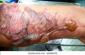
• Second degree (partial thickness) burn: This involves part of dermis. It manifests with blisters, oedema, moist surface and pain (because of the intact sensory nerve receptors) at the affected site. Healing occurs in about 2 weeks, scarring is minimal.
- Third degree (full thickness) burn: Involves complete burn of the dermis. The burned skin looks charred, white or grayish and the surface is pain free.
•Fourth degree: Involves deeper tissue than the skin.
DETERMINING THE BURN EXTENT
The total body surface area (TBSA) burned is calculated using one of several techniques. When calculating TBSA, include those areas of partial and full thickness burns, superficial burns are not included in the calculation
• The Wallace rule of nines (9’s) is the best known method of estimating burn extent.
• Rule of palm- A second technique of estimating TBSA is using the patient’s hand. The patient’s hand represents about 1% TBSA and the total burn size can be estimated by determining how much of the patient’s (not the examiner’s) hand areas are burned
• Lund and Browder charts - more accurate method of assessing burn extent., provide an age-based diagram to assist in precise calculation.
Indications for admission
1. Burns covering >10-15% of total body surface area
2. Burns associated with smoke inhalation
3. Burns involving hands, feet, face, neck, perineum and joint surfaces
4. Burns associated with suspected child abuse
5. Age < 2 years
6. Associated trauma
7. Burns cannot be adequately managed at home
Management
- First aid/pre hospital care:
- Stop the burning process by removing clothing and irrigating the burns
- Extinguish flames by allowing patients to roll on ground, applying blanket, water or other fire extinguishing liquids
- Use cool running water to reduce temperature of the burn
- Chemical burns- irrigate with large volumes of water
- Wrap patient in a clean cloth and transport to the nearest health facility
2. Hospital care:
-rapid assessment: take a quick history( cause of burn, time of injury, area of body involved, circumstances surrounding the burn injury, associated injuries) and physical examination. Note the pattern of injury & plausibility of history, differentiate accidental from Inflicted burns.
-investigations: take blood samples for investigations like PCV, E/U/CR, grouping and cross-matching of blood, urine samples for urinalysis, wound swab m/c/s.
-Resuscitation
- Assess airway-
- Every severely burned patient should receive 100% oxygen
- Assess ventilation
- Assess circulation
- Secure intravenous access
- Pass urinary catheter
- Pass NGT
-fluid therapy:
Parkland formula- 4mls/kg/Burn surface area + maintenance fluid
- ½ of the total volume to be given over 8hrs from the time of injury
- ½ over the rest 16 hours
fluid of choice- ringer’s lactate or plasma.
- Care of the wound:
- Clean with large volume of lukewarm sterile saline
- Gently cover burn with clean linen in 2nd degree burns
- Keep small burns in cool water or paint skin with gentian violet (GV) until pain subsides
- Apply Topical Agents: silver Nitrite, silver sulfodoxine, mafenides
- Tetanus Prophylaxis
- Surgery: debrimo, escharectomy, skin grafting, wound dressings (sulphratule, dermasin)
- Pain management
-antibiotics
-mental/psychological treatment.
Complications.
IMMEDIATE (1st 24 hrs)
- Fluid / blood loss: dehydration, shock, hypotension, electrolyte imbalance, aneamia
- Sepsis
- Hypothermia,
- severe pain,
- Oliguria
- Acute renal failure
LATE (>24hrs)
- Infections: Septicemia, endotoxaemia
- RS: Pneumonia, Pulmonary Embolism
- CVS: Endocarditis, suppurative thrombophlebitis,
- UGS: Urinary tract infection
- MSS: Scarring, contractures, keloids, vitiligo, alopecia
- GIT: curling’s ulcer.
- Psychosocial: cosmetic problems, low self esteem

